Cytoskeleton and Cell Motility
V.1 Coordinators
V.2 Participants
V.3 Introduction
V.4 Specific Research Objectives
V.5 Background and Significance
V.6 Research Plan
- V.6.ii Subproject 2 - Microtubule Interactions with the Cortex
- V.6.ii.a Advantages of C. elegans Model
- V.6.ii.b Aims
- V.6.ii.c Methodology
- V.6.ii.c.1 Identification of regulators of astral microtubule dynamics and interactions
- V.6.ii.c.2 Develop approaches for quantifying astral microtubule plus-end behavior and force generation; and measure changes in those parameters as a function of position in the embryo, stage of mitosis, and presence vs. absence of selected proteins
- V.6.ii.c.3 Mathematical models of rotational alignment, spindle asymmetry, and spindle rocking, based on measurements and predictions
V.8 Timeline
< Previous | Page 7 of 23 | Next >
V.6.ii Subproject 2 - Microtubule Interactions with the Cortex:
How do C. elegans embryos control placement of the mitotic spindle and hence of cleavage planes? We seek to understand how the distributions, dynamics, and interactions of astral microtubules are regulated to achieve the movements of the spindle poles that ultimately lead to the generation of daughter cells of the correct size, in the correct positions, and containing the correct complement of chromosomes and cellular components. We also wish to model cleavage plane specification, cytokinesis, and polarity establishment in C. elegans. Our basic premise is that these processes involve the deployment and spatial regulation of microtubules and various motor molecules. Computer simulations can reveal whether a particular model or class of models is capable in principle of describing a phenomenon, enabling elimination of otherwise plausible models. They will also enable iterative experimentation and refinement toward the goal of predictive models.
V.6.ii.a Advantages of C. elegans Model:The one-cell C. elegans embryo is ideal for investigating regulation of astral microtubules. The embryo is relatively large (~30 x 50 um) and transparent. Recent advances in generating transgenic animals that express GFP-tagged β-tubulin, γ-tubulin, and histones allow us to monitor the dynamic behavior in living embryos of microtubules, spindle poles, and chromosomes, respectively (Strome et al., 2001). Analyzing the consequences of genetic mutation or RNAi depletion of gene products can reveal the functions of microtubule-associated and cortical components. Most importantly, the embryo displays a stereotypical progression of spindle movements that reflect astral microtubule functions and interactions:
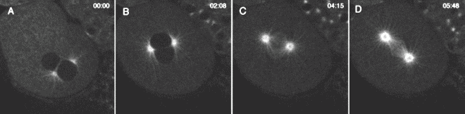
Rotational alignment (Figure 5 A-D). The nascent spindle in the single-celled C. elegans embryo rotates ~90°, which aligns the spindle along the axis of polarity of the cell (anterior-posterior axis of the embryo). This rotation ensures that the axis of division is aligned with the axis of segregation of cytoplasmic, cortical, and membrane components.
|
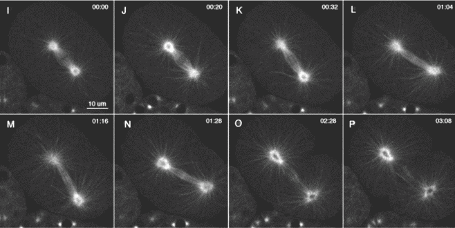
Figure V.5. The dynamics of microtubules and mitosis in a 1-cell C. elegans embryo. Anterior is to the upper left and posterior is to the lower right. (A-C) GFP-β-Tubulin shows paired male and female pronuclei, with 2 associated centrosomes and astral microtubule arrays. The centrosome pair rotates 90° as the nuclei migrate toward the center of the embryo and set up the metaphase spindle. Spindle asymmetry (Figure 5 I-P). During anaphase, the posterior spindle pole moves toward to the posterior cortex, while the anterior spindle pole stays relatively stationary. Because the spindle specifies the placement of the cleavage furrow, the differential movement of the spindle poles leads to an unequal first division, generating a smaller posterior daughter cell. Spindle rocking (Figure 5 I-P). Toward the end of anaphase, the posterior pole of the spindle undergoes dramatic rocking from side to side. Although our preliminary evidence suggests that rocking is not required for spindle asymmetry, rocking provides clear evidence for and an opportunity to investigate the distinctive force-producing interactions of astral microtubules and cortical sites that are different at the posterior and anterior cortex. |
|
Fig. V.5. (continued) A second C. elegans embryo expressing GFP-β-Tubulin showing the dynamic behaviors of the spindle and astral microtubules during anaphase. |